Neighborhood watch in tissues
Max Planck researchers investigate mechanisms controlling macrophage network movement in tissues
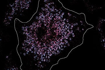
Macrophages are immune cells in the body that remove dead cells and microbes. Therefore, they form large networks of many individual cells in the tissues of all organs, scanning for signs of infection and eliminating pathogens. However, how exactly macrophages move to support their surveillance function in tissues has remained largely unknown. Researchers at the Max Planck Institute of Immunobiology and Epigenetics in Freiburg now show that integrin receptor-mediated migration is crucial for effective tissue surveillance. Surveying cells move forward by grabbing hold of their immediate surroundings using the integrin receptor proteins. Without this receptor functionality, the macrophages were severely impaired in their movement, and they were not able to form an effective surveillance network to pick up particles and dead cells.


Immune cells populate practically all our organs to protect our body and maintain it in a healthy state. Some immune cells sit throughout their lifetime in tissues in a constant surveillance mode. In situations of severe tissue injury or infection with microbes, these cells then receive additional help from immune cells recruited from the bloodstream.
The lab of Tim Lämmermann at the Max Planck Institute of Immunobiology and Epigenetics in Freiburg investigates the cellular mechanisms controlling immune cells when they maneuver through the spaces of complex tissue environments. In their latest study, the lab set out to answer how macrophages, which form large surveillance networks in tissues, move and coordinate their surveillance activities together to protect our body.
Keep watch in tissues
Macrophages are famous for engulfing and digesting all kinds of debris. They act as scavengers that constantly remove dying cells in developing and adult tissue. When microbes invade our body during an infection, they also alert the immune system and recruit other immune cells for help. Moreover, macrophages can take up and eliminate microbes and foreign particles that have entered our bodies.
“While the different phases of corpse recognition, uptake, and digestion are molecularly well described when it comes to individually responding cells, we only have a relatively limited understanding of how group dynamics regulate these processes. We found this lack of knowledge rather surprising since it has been known for decades that tissue surveillance is a group effort performed by large populations of macrophages, which distribute in tissues as networks comprised of many individual cells. Therefore, we wanted to learn about how individual macrophages move in tissues and which factors influence the dynamic behaviors of whole macrophage networks. By doing so we sought to identify processes that determine the efficiency of macrophage networks in detecting and removing particles as well as dead cells,” says Tim Lämmermann.
To realize these research goals, the Lämmermann team studied the movement of macrophages in artificial three-dimensional environments and mouse tissues. The composition and architecture of mouse tissues is very comparable to human tissues, which is why studies in mice teach the scientists a lot about the processes in humans.
Migration modes of immune cells
“When we talk about macrophages moving around in murine or human tissues, we should not think of them having an unfettered ability to move. Instead, we must appreciate that cells really must make some effort and employ many different migratory processes to move. Productive movement generally entails cells squeezing their cell bodies between other cell types in a tissue as well as through the extracellular matrix, which is a dense scaffold of macromolecules including collagens and other proteins,” says Neil Paterson, first author of the study.
For most immune cell types the preferred form of movement is akin to that performed by amoeba. This means that they can generate propulsion primarily by coordinated changes in their cell shape. To do this they rapidly protrude and retract extensions using cytoskeletal forces. In this way, many immune cells squeeze themselves through tissues, guided by chemical attractants to reach, for instance, sites of tissue wounding. “To our surprise, we found that macrophages that randomly explore tissue spaces do not entirely follow this canonical concept of 3D immune cell migration. Instead, they critically depend on strong adhesive interactions with their tissue environment when they move,” says Tim Lämmermann.
Surveying macrophage networks cannot compensate integrin receptor loss
In this second mode of movement, cells use so-called integrin receptors for force transduction. Thus, surveying macrophages migrate by pulling with these adhesion receptorson on their immediate surrounding such as extracellular matrix fibers. The results by the Lämmermann Lab show that the integrin-mediated movement is critical, especially for the removal of dead cells by macrophage networks. A loss of integrin functionality results in severely impaired particle uptake by a surveying macrophage network.
The work sheds new light on the largely neglected population aspect of tissue surveillance by macrophages. It shows how the dynamics of individual cells in a macrophage network shape the efficiency in tissue surveillance and clearance of dead cells. Together, the results of the Freiburg researchers led by Tim Lämmermann provided new insights into how immune cells maintain tissue health and prevent inflammation.
TL/NP/MR