Quantum leap on film
An ultra-fast microscope combines atomic spatial and temporal resolution and thus enables unprecedented insights into the dynamics of electrons in molecules
In order to better understand (and possibly control) fast chemical reactions, it is necessary to study the behaviour of electrons as precisely as possible – in both space and time. However, up to now, microscopy methods have delivered only either spatially or temporally sharp images. By cleverly combining established techniques of tunnelling microscopy and laser spectroscopy, a team led by Klaus Kern, Director at the Max Planck Institute for Solid State Research in Stuttgart, has now overcome these obstacles. Using their atomic quantum microscope, they can make the movement of electrons in individual molecules visible.
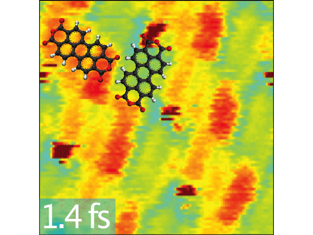
. After about 1.4 femtoseconds (three images), the electron once again reaches the lower-energy state.")
It is essential not only for understanding biological processes (e.g. plant photosynthesis) to map the electron dynamics in molecules but also for many technical applications such as the development of solar cells or new types of electronic components. Until now, imaging methods have sometimes delivered images that are difficult to reproduce – or even contradictory. This is because they cannot map the fast electrons directly but rather must resort to techniques that can only reconstruct the behaviour of the electrons.
Although modern microscopy techniques offer almost unlimited possibilities, certain compromises must be made. For example, scanning tunnelling microscopy with a resolution of one tenth of a picometre (1 × 10⁻¹² m) allows extremely sharp images of individual atoms to be taken. However, this is slow and cannot capture the electron dynamics in a material. On the other hand, optical methods with ultra-fast laser pulses can detect electron movements in the attosecond (1 × 10⁻¹⁸ of a second) range. However, they can provide only coarse, washed-out spatial images – far removed from the atomic resolution possible with scanning tunnelling microscopes. The typical electron dynamics and laser pulses are in the range of a few hundred attoseconds.
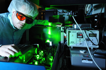
Camera made of scanning tunnelling microscope and attosecond pulses
 at the beginning of the image. The brighter the colour, the higher the electron density: The electrons therefore move back and forth between the centre of the molecules and their edges.")

“For the past few years, we have been working on combining these two techniques in such a way that we can use their strengths without introducing their weaknesses”, says Manish Garg, research group leader at the Max Planck Institute for Solid State Research. To do this, the researchers had to couple tried and tested scanning tunnelling microscopy with state-of-the-art laser technology. In a scanning tunnelling microscope, a wafer-thin, atomically thin tip travels just above a conductive surface. Thanks to the quantum-physical tunnel effect, electrons can flow between the surface and the microscope tip – even if there is no direct contact. For example, a molecule on a surface can gradually be shaved off atom by atom.
The new microscopy technique uses laser pulses in order to modulate the tunnel current by selectively exciting the electrons in the material. “This must be done extremely quickly. Otherwise, thermal effects come into play and make the measurements impossible”, explains Alberto Martin-Jimenez, who played a key role in the experiments. Unfortunately, the necessary ultra-fast laser pulses in the attosecond range are not available “out of the box”. However, thanks to the rapid development of laser technology in recent years, researchers have now been able to generate precisely the right pulses. Two years ago, Garg and Kern demonstrated the function of such an atomic quantum microscope for the first time.
New perspective on biophysical processes, solar cells, and transistors
They have now been able to directly observe electron movement in molecules with this one-of-a-kind instrument. With the help of the ultra-short pulses, the electrons in the molecule can be excited to jump between the different orbitals. This was noticeable in the tunnel flow. The highlight of the new technique is being able to fire two minimally time-delayed pulses in quick succession at the molecule to be investigated with an exact time interval and scan it in the process. If this procedure is repeated several times and the time interval between the pulses varied, an image series that reproduces the behaviour of the electrons in this molecule with atomic accuracy is obtained. The fast laser pulses thus provide the information about the electron dynamics whilst the scanning tunnelling microscope precisely scans the molecule.
“This allowed us to directly map the dynamics of electrons in molecules – how they jump from one orbital to another – for the first time”, says Garg. “This basic technology provides completely new possibilities for directly observing quantum mechanical processes such as charge transfer in individual molecules and thus better understanding them”. It is still not possible to predict the possible areas of application for such a quantum microscope. Especially in charge transfer processes, which play a crucial role in many biophysical reactions as well as in solar cells and transistors, it could provide crucial new insights.