New insights in brain tomography
Functional imaging signals indicate weakening in brain activity
Neuroscientists use functional neuroimaging for everything from understanding how the brain thinks to investigating illnesses. Recently it has also helped to diagnose neurological and psychological disturbances - and even to plan neurosurgery. Researchers from the Max Planck Institute for Biological Cybernetics in Tübingen have now published a paper in Nature Neuroscience which shows that impaired functional neuroimaging signals - considered "negative" compared to normal ones - are connected to a reduction in neuronal activity. Knowing this gives scientists even broader uses for functional neuroimaging.
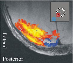
 and positive (red) answer the visual stimuli. Insert: The blue square shows the part of the visual area that was not stimulated, the red square the part that was stimulated.")
Different parts of the brain are active during different kinds of activity: perceiving an object, for example, or solving equations. These parts also have a specific effect on brain metabolism and blood supply. For example, if you are given a recognition task, more blood goes to areas that are specifically active during the visual recognition process. Because the blood flow momentarily increases in active brain areas, the oxygen concentration also goes up there. This principle allows for the study of brain activity connected to a number of different tasks and conditions; the field is known as functional neuroimaging.
Most studies within functional neuroimaging use techniques like magnetic resonance imaging (MRI), position emissions tomography (PET), or optical imaging. Using these techniques, it is possible to measure spatial and temporal changes to the blood flow or oxygen concentration in the brain. They measure what are essentially secondary signals; the actual neuronal electrical activity is not seen. In order to interpret functional neuroimaging signals properly, scientists must understand the relationship between the signals and the neuronal activity behind them.
Besides the usual increases in signal strength, neuroimaging studies also occasionally show prolonged reductions in blood flow and the brain's blood oxygen levels. These relatively "negative" signals can indicate vascular processes, and be independent of local changes in neuronal activity. Increased blood flow in one area can cause a reduction in other areas, because the total amount of blood is limited. Scientists call this the "steal effect". If this scenario is accurate, these negative signals can be categorised as epiphenomena. However, it may be inaccurate - and the negative signals could also reflect a reduction of neuronal activity.
In a study recently published in Nature Neuroscience, scientists from the Max Planck Institute for Biological Cybernetics in Tübingen investigated the connection between reductions in functional MRI signals and the neuronal activity that causes them. They used a sensory stimulus which only stimulates parts of the visual field. The regions in the rear part of the brain responsible for processing visual information are organised into maps where small groups of neurons represent parts of the visual field. If the scientists stimulate only one part of the visual field, this causes negative signals in the regions that do not represent the simulated part.
Amir Shmuel, Mark Augath, Axel Oeltermann and Nikos Logothetis discovered that this negative functional imaging signal is in fact connected to a reduction in neuronal activity. In synapses, and in the bodies of local neuron cells, electrical activity decreased. The dropping in synaptic activity reflects a reduction in input in local neurons, and dropping action potentials - produced by cell bodies - also lead to a reduction in output signal. Because a reduction in neuronal activity immediately followed, the later blood flow decrease could not have been the reason for it. The reduction in neural activity could not have been caused by a "steal effect".
The scientists consider it most likely that a significant part of functional MRI negative signals are triggered by a reduction in neuronal activity. Nikos Logothetis explains, "the study makes it possible to use negative signals in functional imaging to deduce weakening brain activity connected to particular cognitive tasks or neurological illnesses."