The anatomy of memory
Scientists discover new mnemomic networks in the brain
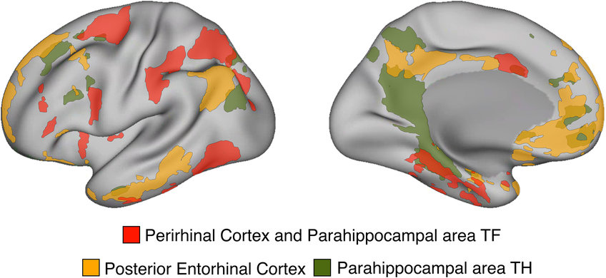
How can the amazing abilities of our memory be explained on the basis of brain anatomy? It is known that different brain functions are anchored in different areas and structures of the brain. For example, we know that certain areas of the cerebral cortex are responsible for perception of the outer world, imagining our future, and thinking about other people. However, little is knowing about connection of the brain regions supporting these important cognitive functions with the human memory system. Using a novel approach of precision neuroimaging and high-resolution functional magnetic resonance imaging (fMRI), neuroscientists and physicists at the Max Planck Institute for Human Cognitive and Brain Sciences in Leipzig (Germany) and anatomist Menno Witter from the Kavli Institute for Systems Neuroscience in Trondheim (Norway) have now ventured into the depths of the human memory system. They discovered previously unknown cortical networks and shed light on the anatomical organization of the human memory system.

The human memory system is seated in the medial temporal lobe (MTL). Broadly, it contains the hippocampus, parahippocampal cortex, perirhinal cortex, and entorhinal cortex. “One big challenge in studying the MTL is its great anatomical variability across people. Therefore, prior studies that were using group-averaged data, blurred fine anatomical details between different subregions of the human MTL that are located in close proximity to each other. It is like studying face structure by averaging 1000 different faces together. We will get important organizational principles of a face – where the eyes and the nose are located, where the mouth is, but we will completely loose idiosyncratic important details”, explains the study's first author, Daniel Reznik of the Max Planck Institute for Human Cognitive and Brain Sciences, Leipzig. According to him another challenge in studying the MTL in humans is that this brain region is strongly affected by susceptibility artefacts, therefore the ability to get good-quality signal from this brain region is highly limited.
In the current study, the scientists solved these challenges in MTL imaging and finally explored the distributed cortical anatomy associated with different subregions of the human temporal lobe in individuals.
New cortical pathways in the human memory system
Exploring the depths of the human memory system
“So instead of collecting data from many different people, we collected a lot of data from the same individuals, which dramatically increased the anatomical precision of our study. We combined our expertise in high-field imaging, neuroanatomy, and cognitive neuroscience and examined MTL anatomy in great detail. This allowed us to identify cortical networks associated with the human medial temporal lobe that were unknown to previous human memory research”, Daniel Reznik concludes and adds: “Similar cortical networks also exist in animals and perhaps the most exciting finding is that we have now evidence for potentially new cortical pathways in the human memory system compared with non-human primates."
Christian Doeller, Director of the Department of Psychology at the Max Planck Institute, adds, "These new findings are important since even after many years of research into human memory, no one really knew how the regions in the MTL are connected with the rest of the human brain. Connectivity of the entorhinal cortex is of particular interest for us, since this is one of the first brain regions affected by Alzheimer’s disease. Our discovery defines the anatomical constraints within which human memory functions operate and are informative for studying the evolutionary development of temporal lobe circuitry in different species. For example, data from non-human primates show only slight connections between the entorhinal cortex and the frontal cortex in comparison - in contrast, we found that these connections are more pronounced in humans.”
Daniel Reznik adds: “Since one of the networks connected to the human entorhinal cortex is also involved in social processing, we suspect that it is an evolutionarily young network that may have evolved after the extensive expansion of the cortex in humans."


