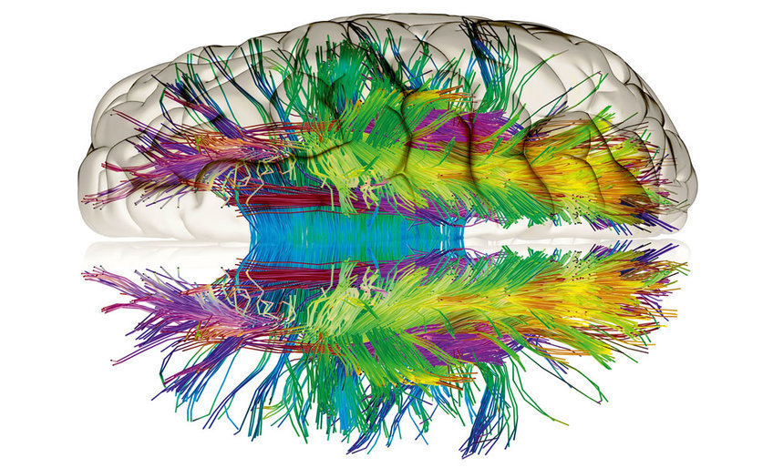
The brain
The human brain is the most complicated organ that nature has ever created: 100 billion nerve cells and many more contact points between them provide our brain with capabilities that no supercomputer can match to this day. One of its most important characteristics is its ability to learn. But how can a collection of neurons learn anything in the first place? And can this ability be specifically improved?
Until a few years ago, scientists thought one thing was certain: an adult’s brain will not change. Today, however, we know that the brain is being constantly transformed right up until old age. Some neurobiologists even draw comparisons with a muscle which can be trained. Sellers of so-called brain jogging programmes are now picking up on this idea, offering exercises which are intended to increase learning and memory performance.
The idea that the brain remains capable of learning for a lifetime remains undisputed from a scientific viewpoint. If it were not, humans would not be able to overcome the wide range of challenges that we encounter during the course of our lives. For example, even in old age we can learn a foreign language and yoga, can remember the face and voice of a new work colleague, or the route to a new pizzeria.
However, many scientists doubt whether brain jogging exercises increase the general performance of the brain. They assume that the effects of the coaching have an impact only on the task which is being trained. According to this view, other capabilities accrue little or no benefit from brain jogging programmes.
News
Synaptic plasticity
What happens exactly when our brain learns and stores something new?
Learning takes place at the synapses – those are the locations at which the electric signals are relayed from one nerve cell to another. Neuroscientists have discovered that synapses can vary the effectiveness of the transfer. This phenomenon is also described as synaptic plasticity. For example, a synapsis can also be strengthened by a procedure called long-term potentiation (LTP), in which the synapsis distributes more neurotransmitters or forms more neurotransmitter receptors. Similarly, the signal transmission at a synapsis can be reduced by long-term depression (LTD).
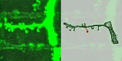
The transmission of signals can, however, not just be strengthened or weakened; it can also be enabled in the first place or completely stopped. For example, neuroscientists know today that synapses can be completely re-formed or dismantled, even in the adult brain. In a few places such as in the olfactory system, new nerve cells can be formed one’s entire life. It is therefore not an exaggeration to claim that our brain is a life-long construction site.
Consolidation and deterioration, building-up and reduction - the strength with which the signals are relayed between nerve cells is being constantly adjusted. A simplified way of looking at it is imagining that the transmission of signals is strengthened when the brain stores something – and is weakened when it forgets something. Many neuroscientists today share the view that the synaptic plasticity is the basis of learning and memory.
Without plasticity, the brain would be missing something fundamental: its ability to learn. Learning is much like sports: the more that a certain skill is needed, the more effective it is handled. If you drive a taxi, for example, you need to have a good sense of direction and remember routes. Your spatial memory will improve as a result of your work each day. This will leave behind traces in the brain, such as in the brain of London taxi drivers: researchers have found that the hippocampus in their brain – an area in the brain which is crucial for spatial memory – will become bigger over time. Clearly a spatial sense of direction being trained in such a way needs more space! It is still unknown whether taxi drivers generally have a better memory.
The plasticity also helps the brain to repair damage at least partially. If nerve cells die off in a stroke, neighbouring areas of the brain can partially take over the functions of the affected area. At the Max Planck Institute for Human Cognitive and Brain Sciences, researchers have found that the brain can in this way partially compensate for damage after a stroke. Scientists at a number of Max Planck Institutes are investigating how the brain and its nerve cells remain plastic.
Another important field of research lies in the connections of the brain. Each of the approx. 100 billion nerve cells in the human brain receives signals from other cells via tree-like structures – which are called dendrites – and offsets them against each other. In doing so, they form their own electric signal that the cell relays to another using a thread-like axon.
Structure of the brain
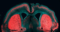
The human brain can be subdivided according to various criteria. It can be explained in terms of evolution, as, like all vertebrates, it consists of an end brain, interbrain, midbrain, hindbrain and medullary brain. Anatomically, the areas known as the cerebrum, interbrain and cerebellum, as well as the brain stem, are particularly noticeable. Particularly striking is the cerebral cortex, which forms part of the end brain. During evolution, it has grown so strongly that it surrounds almost the entire brain. With its ridges and coils, the cortex gives the brain its walnut-like look.
The cerebral cortex is also the home of advanced mental abilities. Individual areas have different tasks in the process. For example, some sections are specialised in understanding language, recognising faces or storing memories. However, normally no single region is responsible for a certain skill, as it works only in concert with others. In the cerebral cortex, both neighbouring nerve cells are linked together (local network) as well as with cells in regions distant from each other (global network).
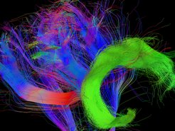
Scientists can examine which areas of the brain are linked with each other with the aid of magnetic resonance imaging (MRT). With this technology, they can make visible the nerve cell structures, grouped together in fibre strands, which connect the areas of the cerebral cortex. By following this procedure, linguists discovered an area of the brain crucial for language abilities: the Fasciculus Articuatus. Without this bundle of nerve fibres, toddlers could not form and understand complex sentences. This is possible only once this connection has sufficiently developed. In apes, by contrast, these nerve fibres remain poorly developed for their entire life. As a result, these animals will not manage, even after years of training, to form even the simplest of sentences – despite the presence of other obligatory brain areas and the anatomical conditions for speaking.
With a variation of this technology – so-called functional magnetic resonance imaging - scientists can distinguish between active and non-active regions of the brain. In doing so, they have learned much about the structure and functionality of the brain. For instance, Max Planck researchers from Leipzig found out why an imbalance between brain activity of the left-hand and right-hand side of the cerebrum occurs in people who stutter: Within the overactive right-hand network, they discovered a fibre tract that is significantly more pronounced than with people without language problems. The stronger the so-called frontal aslant tract is, the more strongly a person will stutter.
MRT technology cannot be used, however, to make an exact map of the circuitry of the brain, as the precision of the method is not high enough. After all, up to 10,000 synapses are present in one nerve cell; the total number is 100 trillion. That shows how dense the communication network in the brain is. In this network, neighbouring nerve cells can be linked to each other, but also cells which are far away from each other. Untangling this jumble of local and global connections – also known as a connectome – is the goal of the Max Planck researchers.
The connectome – wiring diagramm of the brain
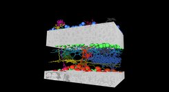
For this reason, the scientists are developing new methods to help them decipher the connectome. Their models for this are mice: recently, they examined the circuitry of areas in the retina as well as in the cerebral cortex and found that nerve cells in the so-called entorhinal cortex are organised like a transistor: before a nerve cell can activate another cell, it contacts an impeding cell and its own activity is therefore inhibited.
Using these kinds of circuit diagrams, scientists want to learn how the brain works. At Max Planck Institutes, they are already working to explain the principles of information processing. Currently, they are focusing on brains that have a simpler structure and contain fewer nerve cells and fibres than the human brain. Mice are one such model case for neuroscientists. As mammals, they have a brain which is structured and functions similarly to a human. By using it, researchers can investigate everything from the fundamental functioning of nerve cells to the circuitry within the cerebral cortex.
The brains of zebra fish and their larvae have an even more straightforward structure and are therefore easier to examine. The brain of a fish larva, for example, does not just have only 100,000 nerve cells – a million times fewer than a human – it is also almost completely transparent. Scientists are therefore able to look into the interior of the brain with their microscopes without a surgical procedure.
Invertebrates can also serve as a model for neuroscientists. Their nerve cells are admittedly very small, making their activity harder to measure. But because of their relatively more straightforward architecture, the principles of interconnections used for perceiving and processing external stimuli can be analysed. For instance, researchers can learn from the brains of fruit flies how hunger influences decision-making processes. By analysing the vision system of blow flies, they want to discover how the insects can perceive movements so incredibly quickly. Even a very simply structured organism like the threadworm C. elegans can deliver important insights into the development and operation of the nervous system.


