Head-mounted microscope measures neuron activity
Miniature device enables scientist to record nerve cell activity in all cortical layers in lit environments
Researchers of the Max Planck Institute for Neurobiology of Behavior have developed a miniature microscope small enough to be carried on the head of freely a moving mouse and capable of measuring neuronal activity in all cortical layers, even the deepest ones. The two-gram microscope can be controlled remotely, which minimizes the need to handle the animal. The microscope also incorporates new technology enabling imaging in lit environments, something that all comparable microscopes struggled to do. Neuronal activity can now be imaged from all cortical layers in the freely moving mouse during the full range of the animal’s behaviors. This new microscope is a game changer for exploring the link between neural activity and complex animal behavior.


How does the brain produce behavior? Studying the brain during behavior is most informative when the animal is free to interact with its environment in any way it chooses. This requires small head-mounted devices that provide access to the brain, but that do not interfere with the animal’s behavior.
“We are interested in how freely behaving animals use vision to make decisions in their everyday life. As many of the brain cells that are thought to be involved in this process are located deep in the visual cortex, we made a very light-weight head-mounted microscope that can measure activity from these neurons but does not interfere with the animal’s behavior. We have made a large step towards finally imaging brain activity deep in the cortex of animals performing natural visual-based behaviors” says Jason Kerr, director of the Department of Behavior and Brain Organization, Max Planck Institute for Neurobiology of Behavior in, Bonn, Germany.
Imaging from all cortical layers
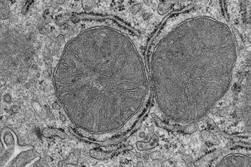
In the study the researchers developed a miniature 2-gram three-photon excitation microscope which delivers a number of firsts. For the first time, it is now possible to image neural activity on a single-cell level from all cortical layers without having to interfere with the animal’s behavior, made possible by a remote focusing mechanism. Its modular design also offers a high-resolution configuration which allows for functional recordings from neuronal somata and dendrites. Another important feature is that, due to a modified detector system, the microscope can be used in fully lit conditions. “The robustness of our new miniature microscope to ambient light allows us to image the brain’s activity while the animal is having access to its full sensory repertoire. We can now study visually guided natural behavior like prey hunting and predator avoidance” says Alexandr Klioutchnikov, first author of the study.
To confirm range and stability of the new miniature three-photon microscope, the team imaged from the deep cortical layers 4 (L4) and 6 (L6) while mice were freely exploring an arena. It turned out that L4 and L6 neurons were differentially modulated depending on the environmental light. As the microscope can easily be mounted again at the exact same position, the same neuronal populations can be imaged in follow-up sessions spread over days. This opens up the possibility to monitor changes in the brain’s activity, for example while the animal is learning.