Understanding the human brain
Functional magnetic resonance images reflect input signals of nerve cells
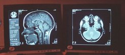
The development of magnetic resonance imaging (MRI) is a success story for basic research. Today medical diagnostics would be inconceivable without it. But the research took time to reach fruition: it has been nearly half a century since physicists first began their investigations that ultimately led to what became known as nuclear magnetic resonance. In 2001, Nikos K. Logothetis and his colleagues at the Max Planck Institute for Biological Cybernetics in Tübingen devised a new methodological approach that greatly deepened our understanding of the principles of functional MRI.

The great advantage of functional magnetic resonance imaging (fMRI) is that it requires no major interventions in the body. In fMRI, the human body is exposed to the action of electromagnetic waves. As far as we know today, the process is completely harmless, despite the fact that fMRI equipment generates magnetic fields that are about a million times stronger than the natural magnetic field of the earth.
The physical phenomenon underlying fMRI is known as nuclear magnetic resonance, and the path to its discovery was paved with several Nobel prizes. The story begins in the first half of the 20th century with the description of the properties of atoms. The idea of using nuclear magnetic resonance as a diagnostic tool was mooted as early as the 1950s. But the method had to be refined before finally being realised in the form of magnetic resonance imaging.
Today, MRI not only produces images of the inside of our bodies; it also provides information on the functional state of certain tissues. The breakthrough for fMRI came in the 1980s when researchers discovered that MRI can also be used to detect changes in the oxygen saturation of blood, a principle known as BOLD (blood oxygen level dependent) imaging. There is a 20 percent difference between the magnetic sensitivity of oxygenated arterial blood and that of deoxygenated venous blood. Unlike oxygenated haemoglobin, deoxygenated haemoglobin amplifies the strength of a magnetic field in its vicinity. This difference can be seen on an MRI image.
fMRI has given us new insights into the brain, especially in neurobiology. However, the initial phase of euphoria was followed by a wave of scepticism among scientists, who questioned how informative the “coloured images” really are. Although fMRI can in fact generate huge volumes of data, there is often a lack of background information or basic understanding to permit a meaningful interpretation. As a result, there is a yawning gap between fMRI measurements of brain activity and findings in animals based on electrophysiological recordings.
This is due mainly to technical considerations: interactions between the strong MRI field and currents being measured at the electrodes made it impossible to apply the two methods simultaneously to bridge the gap between animal experiments and findings in humans.
fMRT shows input signals
In 2001, Nikos Logothetis and his colleagues at the Max Planck Institute for Biological Cybernetics in Tübingen were the first to overcome this barrier. With the help of special electrodes and sophisticated data processing, they showed unambiguously that BOLD fMRI actually does measure changes in the activity of nerve cells. They also discovered that BOLD signals correlate to the arrival and local processing of data in an area of the brain rather than to output signals that are transmitted to other areas of the brain. Their paper was a milestone in our understanding of MRI and has been cited over 2500 times worldwide.

Their novel experimental setup enabled the Tübingen scientists to study various aspects of nerve cell activity and to distinguish between action potentials and local field potentials. Action potentials are electrical signals that originate from single nerve cells or a relatively small group of nerve cells. They are all-or-nothing signals that occur only if the triggering stimulus exceeds a certain threshold. Action potentials therefore reflect output signals. These signals are detected by electrodes located in the immediate vicinity of the nerve cells. By contrast, local field potentials generate slowly varying electrical potentials that reflect signals entering and being processed in a larger group of nerve cells.
Applying these three methods simultaneously, the Max Planck researchers examined the responses to a visual stimulus in the visual cortex of anaesthetized monkeys. Comparison of the measurements showed that fMRI data relate more to local field potentials than to single-cell and multi-unit potentials. This means that changes in blood oxygen saturation are not necessarily associated with output signals from nerve cells; instead, they reflect the arrival and processing of signals received from other areas of the brain.
Another important discovery the Tübingen researchers made was that, because of the large variability of vascular reactions, BOLD fMRI data have a much lower signal-to-noise ratio than electrophysiological recordings. Because of this, conventional statistical analyses of human fMRI data underestimate the extent of activity in the brain. In other words, the absence of an fMRI signal in an area of the brain does not necessarily mean that no information is being processed there. Doctors need to take this into account when interpreting fMRI data.

