HIV capsid reaches the cell nucleus intactly
Advanced imaging methods provide detailed insights into infection processes
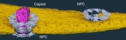
For the first time, scientists at the Center for Integrative Infectious Diseases Research at the University Hospital Heidelberg, the Heidelberg European Molecular Biology Laboratory and the Max Planck Institute of Biophysics in Frankfurt have succeeded in imaging the transport of the HI virus into the nucleus of the infected cell. These electron tomographic images show that the protein envelope of the virus passes through the nuclear pore as a whole and only ruptures inside the cell nucleus, where it releases the viral genetic information. The work thus explains a central mechanism of how viral genetic material is integrated in infected cells.
 passes as a whole through a pore (grey) into the cell nucleus (yellow), where it disintegrates and releases the viral genetic material. (NPC: nuclear pore complex)")
The human immunodeficiency virus (HIV-1) primarily infects certain cells of the immune system and in this way massively weakens the body's own defense against diseases. The genetic material of the virus is securely packaged in a cone-shaped protein capsule, the capsid, which is composed of individual hexagonal parts. While it is known how the capsid enters the interior of the cell through the cell envelope during infection, it has remained unclear how the viral genetic material is passed from the capsid to the cell nucleus, where it triggers the formation of new viruses.
Here, the recently published work provides new insights. Drawing on newly developed methods for the three-dimensional representation of molecular complexes in virus-infected cells, the scientists succeeded in mapping the viral capsid while it was transported into the nucleus. "Until now, it was assumed that the virus capsid does not fit through the pores," explains Hans-Georg Kräusslich, Medical Director of the Center for Integrative Infectious Diseases Research at the University Hospital Heidelberg. "However, the question of how the viral genome enters the cell nucleus is essential for its replication. Our results therefore support the search for new targets in future therapeutic approaches." Although current treatment options can suppress the multiplication of viruses in the body, they cannot eliminate them entirely.
Platforms for high-resolution imaging
For a detailed look at the inner workings of infected immune cells, the scientists relied on high-resolution imaging techniques: With the help of the Electron Microscopy Core Facility at the University of Heidelberg and the Cryo-Electron Microscopy Service Platform at EMBL, they combined various methods of light and electron microscopy and reconstructed three-dimensional images of the molecular structures from the data. In this way, the composition and architecture of the viral complexes and their interaction with the cellular structures in high resolution was visualized. "Our close collaboration and the combination of specialized techniques enabled us to understand yet another aspect of the HIV-infection’s mechanism of action," says Martin Beck, Director and Scientific Member at the Max Planck Institute of Biophysics in Frankfurt since 2019 and former Research Group Leader at EMBL.


