Social Networking at the Cellular Gateway
Researchers reveal nanoscale architecture of cellular uptake machinery
The receptor-based uptake of nutrients and signals, the so-called clathrin-mediated endocytosis, is essential for the growth and communication of cells, but also a common gateway for cytotoxins and viruses. A research team from the Max-Planck Institute for Terrestrial Microbiology in Marburg, German, has now described the functional proximities of the proteins involved for the first time. The results shed new light on this fundamental, yet highly complex, modular and transient transport machinery and contribute to a better understanding of transport processes and their regulation.
.")
All cells, either of a relatively simple yeast microorganism or of a complex human tissue, needs to acquire signals and nutrients from their environment and renew their plasma membranes, which shield them from their surroundings. The primary process essential for these tasks is the clathrin-mediated endocytosis (CME). During endocytosis a small area of the plasma membrane folds inward to form an spherical structure, a so-called endocytic vesicle, for transporting nutrients, signals and membrane into the cell.
Making an endocytic vesicle is a delicate artwork and tremendous hard work at the same time, as the plasma membrane does not bend easily. It is thus not surprising that troops of more than 50 proteins collaborate in this complex machinery. While past studies provided a solid part-list of the “endocytic machinery”, its protein architecture remained poorly understood. However, we need this information to understand how endocytosis works in heath and disease, for example for its eventual manipulation for optimized drug delivery or selective molecular uptake (scavenging toxic or valuable compounds by cells), or to understand how viruses gain cell entry by hijacking the CME.
FRET is used at high protein densities
; scale bar, 2 μm.")
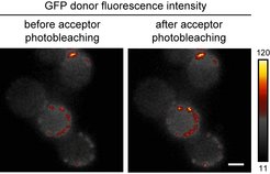
An analysis of the protein organization of the endocytic site is not an easy task, because all the proteins are packed in a very small area (approximately 50 nm radius), which is not accessible for majority of light-microscopy techniques. For researchers of the Max Planck Institute for terrestrial Microbiology in Marburg, the high protein density of the endocytic site was rather an advantage. The group uses FRET microscopy, a high-resolution method, to investigate protein–protein proximities.
The photo-physical phenomenon of Förster resonance energy transfer (FRET) can occur between two fluorescently-labeled proteins only if they are close to each other (typically less than 10 nm), and it can be seen as a loss-of or gain-in-fluorescence of one of them. Detected fluorescence changes can therefore reliable report the protein proximities and map protein neighborhoods inside the cell.
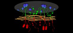
Endocytic proteins form a network
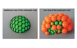
 In the traditional view, coat proteins were though to be buried inside the clathrin cage to connect it to the plasma membrane. (Right) According to our study, key coat proteins span through the clathrin cage to interact with the actin cytoskeleton inside the cell.")
The researchers applied this method to map proximities of 18 core endocytic proteins shared by many organisms, including human. They constructed more than 200 yeast strains containing individual fluorescently labeled protein pairs, of which over 50 pairs showed FRET signals.“This approach allowed us to draw a “social network” inside the endocytic site”, Group leader Michal Skruzny explains. “It was usually thought that the endocytic proteins were randomly placed between the membrane and a so-called clathrin cage. But surprisingly, it is contrary to that: We found the coat organized into several layers with the important endocytic proteins protruding through the clathrin into the cytoplasm. Here they functionally interact with actin filaments that pull the endocytic vesicle inside the cell.”
A newly developed FRET protocol revealed even more: some proteins rearrange in the course of endocytosis. For example, a key endocytic protein that is initially hidden inside the clathrin coat pokes out of it later during endocytosis, when the actin is around.
Methodological framework for multiprotein analysis
Altogether, the research team determined the nanoscale architecture of the core part of the endocytic machinery in detail. Encouraged by their results, they now continue to map the other ever-important protein group of the endocytic site, proteins of WAS/Myo complex. Though these workers come to the endocytic event very late, without their collaborative effort no actin filaments and subsequently no endocytic vesicles can be made. Michal Skruzny concludes: “We believe that our study will greatly contribute to the mechanistic understanding of the endocytic process and its regulation. Furthermore, the FRET microscopy techniques we develop offer a strong methodological framework for characterization of other multiprotein assemblies involved in fundamental cellular processes.”