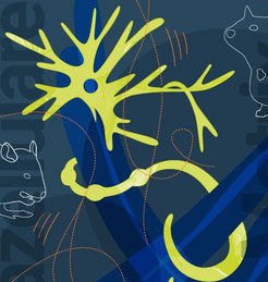
Helping damaged nerves to re-grow
Severed nerve tracts are very difficult to treat. If at all, the damage so far can only be repaired through complex operations. At the Max Planck Institute for Polymer Research, we have developed materials that stimulate damaged nerves into growth. Results from initial tests on mice show that nerve tracts can regenerate this way.
Text: Christopher V. Synatschke / Tanja Weil
Have you ever tried holding a pen without using your thumb? Then you will know how difficult this is. What may seem like an interesting finger exercise is for many a bitter reality. If nerve tracts are damaged or completely severed as a result of a traffic accident or occupational injury, individual limbs or even entire body parts can become numb and often can no longer be moved. In the past, the only chance to restore their functionality has been through surgery. Some operations involve removing nerve strands from another part of the body and reinserting them in the damaged spot. In this way, the damaged nerve endings can grow back together again, restoring a certain degree of movement to the affected part.
Growth requires structure

Although nerves may be able to bridge a severed connection, the process is extremely complex and not always successful. In addition, a framework of proteins surrounds healthy nerves, and injured nerve fibres depend on this framework remaining intact. However, injuries often damage not only the nerve tract itself but also this framework. This so-called extracellular matrix forms the scaffolding for nerve tracts. Just like tomato plants need a trellis, nerve cells need this matrix to grow alongside. At the Max Planck Institute for Polymer Research, we have developed a material consisting of endogenous building blocks, which can be used to replace this matrix. And as was shown, the artificial framework helps the damaged nerves to regenerate themselves. The natural matrix consists of particular proteins: long chain molecules folded like balls of wool. Large numbers of these tiny balls of wool align themselves to form long fibres. These various fibres form a web – the extracellular matrix – that the nerve cells can latch onto.
Lego-build fibres
In order for these proteins to form, numerous complex biochemical processes have to take place within the body – too complex to be recreated in a test tube. Our research takes a different approach: although we use the same basic materials that make up the extracellular matrix, we assemble them in a simpler form. We use short-chain molecules known as peptides, which, like proteins, are composed of amino acid building blocks. We produce these peptides with chemical precision, allowing us to determine the exact position of each individual building block.
To use an analogy, our precise chemical design creates ‘studs’ and corresponding ‘holes’ on the molecules, similar to Lego bricks. Two peptide molecules synthetisised in this way will naturally align themselves so that stud and hole meet. This then creates a stable structure. We were able to use this technique to produce long fibres that – despite their differing microscopic structure – strongly resemble the fibres of the nerve’s extracellular matrix in shape and chemical composition.
From test tube to mouse
How do nerve cells behave when they are to grow on this artificial extracellular matrix? How do these growth characteristics change when we alter the peptides originally used? We investigated these questions in collaboration with our par tner Bernd Knöll, Professor at the Institute of Physiological Che mistry at Ulm University. We produced various peptide structures, deposited them on glass substrates, and cultivated nerve cells on them. While the ner ve cells on some fibr e structures barely grew at all, on others we saw the rapid formation of axons, thin protrusions that create the connections to other nerve cells.
Together with our colleagues at Ulm University, we then used animal models to test the fibre structure that supported the best nerve cell growth. We surgically severed the facial nerve of a mouse on one side, which controls the movement of its whiskers. We then took the fibre-forming peptides and injected them into the gap in the nerve. After 18 days, the mouse was able to move its whiskers again to some extent; the nerve tracts had apparently grown back together.
Since the peptides used our artificial fibres resemble the natural proteins in the extracellular matrix, we are hoping that while the material remains in place during the healing process, the body can then break it down over time. So far we have been able to show that the material remaining at the injection site is slowly decreasing. However, whether this is due to biological degradation or the distribution in the body requires further investigation.
Pioneering properties
As shown by the laboratory experiment in mice, initial damage to nerve tracts can be repaired using our artificial matrix. Before using the material in clinical applications, however, further optimisation is required since the nerve cells on our material do not grow as well yet as they do in the natural matrix. They also grow in a quite disordered manner in all directions. Our next step will be to embed so-called growth factors into the artificial matrix to further accelerate the healing process. Furthermore, we want to orient the injected fibre structures to assist the nerve cells to grow in a specific direction.
We are confident that our artificial extracellular matrix could represent a good alternative to complex surgery for minor injuries to nerve tracts. Further research might also lead to a method of treating not just injuries to the peripheral nervous system but also to the central nervous system.