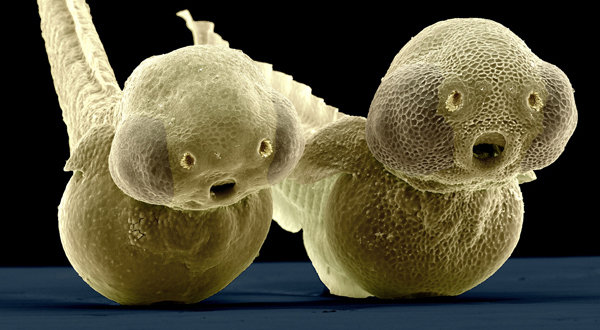
Research on zebrafish at Max Planck Institutes
Scientists in the Max Planck Society carry out research on zebrafish to investigate a number of areas: the development from the ovum to the adult animal, the formation of organs and the functioning of the brain.
Control centre for blood vessels

Researchers at the Max Planck Institute for Heart and Lung Research, for example, have identified the gene in zebrafish DNA that stimulates the growth of blood vessels in the growing embryo. According to their findings, the cloche gene, as it is known, controls the entire development programme for the vascular system. All other genes known to be involved in the production of vessels only become active after the cloche gene is activated. Genetically modified embryos with an inactive gene do not form any blood vessels and die soon after fertilization. Mammals, including humans, have a gene closely related to the cloche gene.
Division of labour in the brain
The relatively simple structure of the nerve system in fish larvae makes it easier for neurobiologists to explain processing operations in the brain. Researchers at the Max Planck Institute of Neurobiology in Martinsried have identified a group of just 15 neurons that control the direction in which the tiny larvae swim. A small region in the hindbrain is responsible for propulsion: it acts like an engine and drives the fish on. In the human brain, a similar division of labour exists between different areas of the brain responsible for movement. Tasks in our brain can presumably be distributed in the same way as they are in the zebrafish. Optogenetics has helped the researchers in this work. They are able to use this technology to modify the fish in such a way that the brain's neurons produce light-sensitive channel proteins. Using short light pulses, the neuroscientists can then open or close the tiny channels to activate the neurons and study their function.
Regenerating the heart
If the heart is not supplied with sufficient blood after a heart attack, heart muscle cells die off. Humans cannot replace this tissue. The opposite is the case for the zebrafish larvae. They can regenerate parts of their heart that have been damaged. Scientists at the Max Planck Institute for Heart and Lung Research in Bad Nauheim have observed in fish larvae with damaged heart muscles that muscle cells from the undamaged atrium migrate into the ventricle and change into ventricular cells. They thus contribute significantly to the regeneration of the heart muscle. If it were possible to stimulate this type of cell transformation using gene therapy, the self-healing powers of the heart could be strengthened.
New blood vessels develop following injury

Scientists at the Max Planck Institute for Molecular Biomedicine in Münster are taking advantage of the surprising regenerative abilities of the zebrafish. Tail fins that have been damaged or clipped off can regenerate the fish completely. It means that all the tissue and cell types can grow back within a few weeks and form a new fin. The necessary blood vessels initially grow from a disordered mesh-like structure into a new fin. The Max Planck researchers have now discovered that some cells from this mesh turn around and migrate against the general direction of growth. These cells then form the arteries at a later stage. The scientists noticed a similar phenomenon in the retinas in the eyes of mice. They have probably thus discovered a common phenomenon of angiogenesis (vascular regeneration). Diseases of the vascular system in humans could be better explained with this knowledge, for example shunts – pathological direct connections between arteries and veins.

